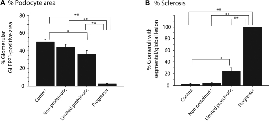
Figure 3.
Quantification of histology: Quantitative information related to Figure 2. (A) Percentage of glomerular tuft area containing GLEPP1 is significantly reduced in progressors and limited proteinurics in relation to controls. (B) Percentage of glomeruli with segmental or global lesions is significantly increased in progressor and limited proteinuric groups compared with control. *P < 0.05 and **P < 0.01 as assessed by Kruskal-Wallis test and then Scheffe test.

