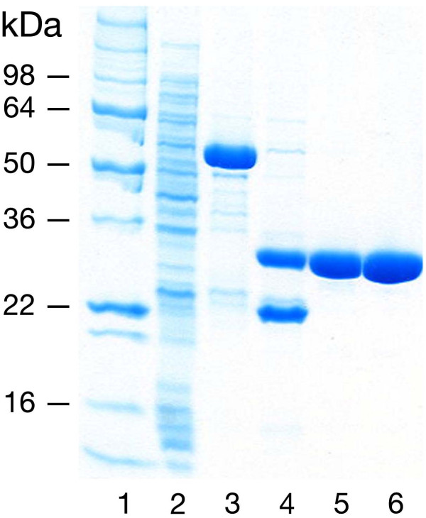
Figure 3.
Representative MK2 purification: MK2(47–366, T222E). Protein fractions were separated by 4–20% SDS-PAGE; stained with Coomassie Blue. Lanes: (1) MW markers; (2) glutathione affinity column flow-thru; (3) glutathione affinity column eluate (40 mM glutathione, pH 8.0); (4) TEV protease cleavage; GST is present below MK2; (5) MonoS 10/10 eluate (~200 mM NaCl); (6) Superdex 75 10/60 peak fraction.

