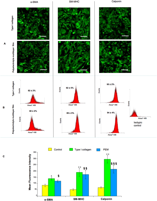
Figure 5. Phenotype stability under normoxia.
After the third passage, the smooth muscle cells phenotype stability of differentiated cell cultivated under normoxic conditions was investigated by confocal microscopy observation (A) and flow cytometry analyses (B, C). A: Microscopical observations show positive cells for contractile markers: α- Smooth Muscle Actin (α-SMA), Smooth Muscle Myosin Heavy Chain (SM-MHC) and Calponin confluence on both coated surfaces (type I collagen and Polyelectrolyte Multilayer films (PEMs)). Objective×40, NA = 0.8, scale bars 75 µm. B: Flow cytometry showed that about 90% cells expressed SMCs markers. C: Mean fluorescence intensity analyses showed a higher SMCs contractile markers expression for differentiated cells compared to control (mature SMCs) whatever the surface coating. (§) PEMs versus control, (*) Collagen versus control. (§ and *: p<0.05, §§ and **: p<0.01, and *** p<0.001).

