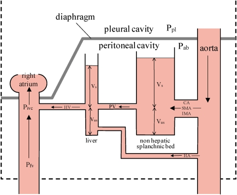
Figure 4. Hydraulic model of the splanchnic vascular bed, consisting of two capacitors, the liver vasculature and the non-hepatic splanchnic vascular bed.
CA = celiac artery; SMA = superior mesenteric artery; IMA = inferior mesenteric artery; HA = hepatic artery; Vs = stressed volume of blod where vascular transmural pressure >0; Vus = unstressed volume where vascular transmural pressure <0; Ppl = pleural pressure; Pab = abdominal pressure; PV = portal vein; HV = hepatic vein; Pfv = pressure in the femoral vein; Pivc = pressure in the inferior vena cava at the entry of the hepatic vein. The height of the column of blood indicates that there is a pressure gradient from aorta > mean vascular pressure in the non hepatic vessels > mean vascular pressure in the hepatic vascular bed > right atrial pressure.

