
Figure 1.
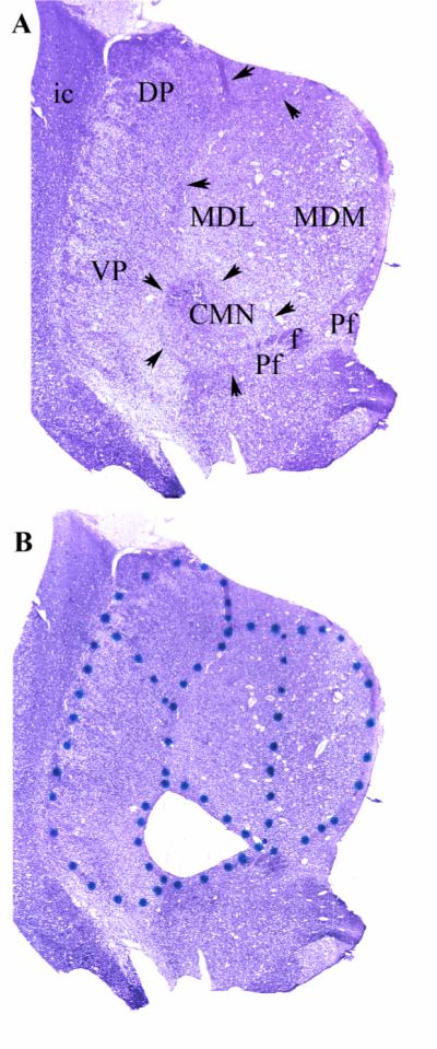
Figure 1: A) Representative uncoverslipped thionin stained 20 micron coronal frozen section through the middle of the thalamus at the level of the CMN. Arrows indicate the internalmedullary lamina which surrounds the CMN and forms the lateral and dorsal borders of the MDN. The ventromedial border of the CMN is reinforced by fascicles of the prominent fasciculus retroflexus (f) and portions of the parafascicular nucleus (Pf). In these preparations the distinction between the parvocelluar and magnocellular divisions of the MD could not be made definitively on the basis of cytoarchitectonic criteria. The nucleus was, therefore, bisected to give medial and lateral halves. Because of the topography of the MD projection to prefrontal cortex, the lateral portion (MDL) projects to the more lateral portion of the prefrontal cortex, while the more medial portion (MDM) projects more medially(Goldman-Rakic and Porrino 1985). MDL and MDM correspond approximately to the parvocellar and magnocellular divisions of MD, respectively. The lateral border of the thalamus is the easily discernible external medullary lamina which intervenes between the reticular nucleus and the internal capsule (ic). The boundary between VP (which contains both VPL and VPM) and DP was made on the basis of neuronal orientation. The neurons of VP exhibit a prominent alignment along diagonally oriented fibers, whereas, the neurons of DP are more homogenously arranged. The medial borders of DP and VP are established by the internal medullary lamina (arrows) that constitutes to lateral border of the MD and CMN.
B) Same section as in panel A after laser microdissection of the CMN. The dots were placed on the glass side of the Leica microdissection slides to delineate the divisions to be dissected (see Methods).