Fig. 3.
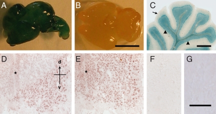
Localization of MISRII in the nervous system. (A–C) MISRII-Cre-lacZ lineage tracing. (A) Whole brain of a MISRII-Cre-lacZ E16 embryo. (B) Littermate control (Misrii+/+, ROSA26-lacZ Cre). (C) Section of the cerebellum of a MISRII-Cre-lacZ adult mouse showing lacZ stain in neurons (arrowheads) but not the white matter (arrow). (D–G) Immunohistochemical detection of MISRII. Sections of lumbar spinal cord of E12 (D) and E14 (E) embryos stained with an anti-MISRII antibody. The asterisk indicates the neuroepithelium, with the dorsal–ventral (d–v) axis indicated. (F) Section of E14 spinal cord stained with control IgG. (G) Section of the cerebellum from a Misrii−/− mouse stained with the anti-MISRII antibody. (Scale bars: B, 10 mm; C, 1 mm; F, 100 μm.)

