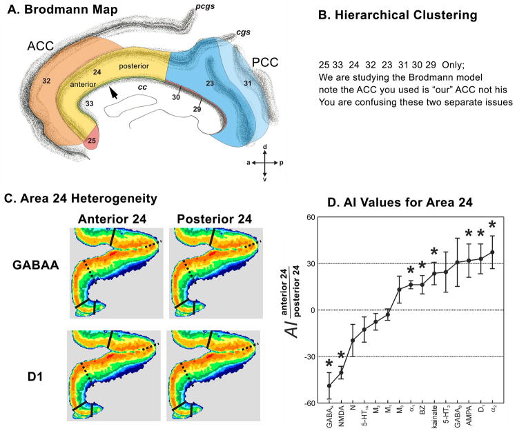
Figure 1.
Flat map of the medial surface of the human anterior cingulate cortex showing the distribution of subregions and areas from a histological assessment. The sulcal depths are noted with a homogeneous gray color. CC: corpus callosum; CG: cingulate gyrus; SCG: superior cingulate gyrus; SFG: superior frontal gyrus; cas: callosal sulcus; cgs: cingulate sulcus; pcgs: paracingulate sulcus.

