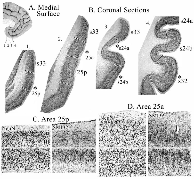
Figure 3.
A. Medial surface of a human hemisphere showing four levels used in B. B. Coronal sections immunoreacted with NeuN antibody through different rostrocaudal levels of sACC. Asterisks indicate the position from which micrographs shown in C, D, and Figure 4 were taken. Sections for SMI32-ir were adjacent to the NeuN sections. C. Micrographs of area 25p show large, densely packed layer II neurons and a very thin layer III D. Micrographs of area 25a showing a wider, but still undifferentiated, layer III. Scale bars = 500 om.

