Figure 6.

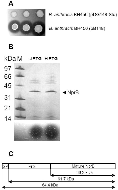
Characterization of B. cereus 569 NprB. (A) 2.5, 5 and 7.5 μl of overnight cultures of B. anthracis BH450 transformed with either pDG148-Stu (upper row) or pB148 (lower row) were spotted on casein agar plates containing 50 μg mL-1 kanamycin. Plates were incubated overnight at 37°C. (B) B. anthracis BH450 (pB148) was cultured in FA broth in the presence of kanamycin (50 μg mL-1) either in the absence or presence of 2 mM IPTG. Supernatants collected at T2 (2 h after the end of exponential growth) were concentrated and applied to SDS-polyacrylamide (10 to 20%) gel using the PhastGel System (Amersham Pharmacia). The gel was stained with Coomassie blue. M designates molecular mass markers. The arrowhead indicates the 38.2-kDa neutral protease B. Results are presented in the upper panel. Protease activities of the corresponding supernatants were determined by spotting them on casein agar (lower panel). The zones of casein proteolysis were observed after 2 h incubation at 37°C. (C) Graphic presentation of the three functional segments of the B. cereus 569 NprB preproenzyme. The protein includes the signal peptide (SP), propeptide region (Pro) and mature protease.
