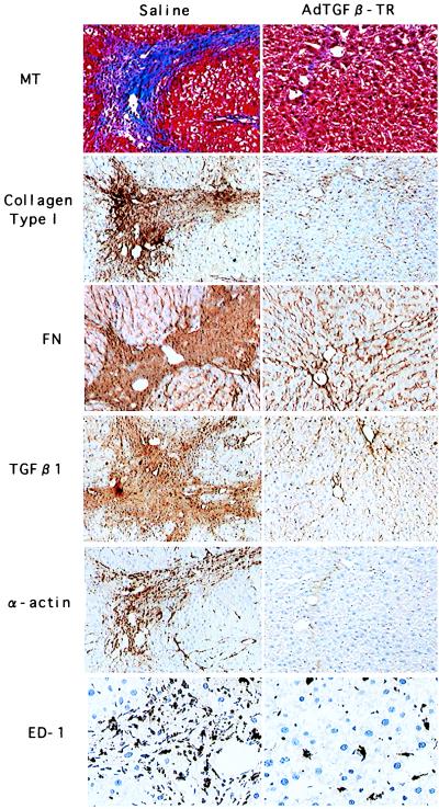
Figure 3.
Immunohistological analysis of liver from rats treated with DMN. Rats were treated as described in the legend to Fig. 1. Liver sections were examined by immunohistostaining by using antibodies against collagen type I, fibronectin (FN), TGF-β1, α-smooth muscle actin (α-actin), or monocyte/macrophage (ED-1). Masson-trichrome staining (MT) also is shown to indicate the fibrotic area. (Left) DMN-treated rats infused with saline. (Right) DMN-treated rats infused with AdCATβ-TR. The upper three sections are serial. [×200 (×400 for ED-1 immunostaining).] Sections from AdCALacZ-infused rats are not shown, because the results were similar to those seen in rats infused with saline.

