Fig. 3.
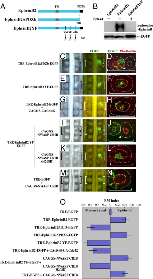
Ephrin cell autonomously coordinates the gap formation and cell epithelialization through repression of Cdc42 activity. (A) A diagram showing mutant forms of EphrinB2. In the mutant EphrinB2YF, three tyrosine residues were replaced by phenylalanines. (B) Western blotting shows that the phosphorylation of EphrinB2 was dependent on interactions between EphA4 expressed in neighboring cells, and also that the phosphorylation was abrogated by the three Y-to-F replacements in the cytoplasmic region of EphrinB2. DF-1 cells that had been separately transfected with EphrinB2 and EphA4 were co-cultured and subjected to Western blotting to detect a phosphorylated form of Ephrin (see Materials and Methods for more details). (C, E, G, I, K, M) Dorsal views of host embryos subjected to a gap-inducing assay as shown in Fig. 2G. DNA plasmids used for the assay are indicated on the left. (D, F, H, J, L, N) Images of horizontal view over a 10-μm thickness obtained by confocal microscopy demonstrate epithelial or mesenchymal states of electroporated cells (green) in a formed somite. Anterior to the left and midline to the bottom. (O) A ratio between the numbers of epithelial and mesenchymal cells that received exogenous DNAs was compared using EM index as previously shown by Nakaya et al. (6). The number of epithelial cells was divided by the total number of electroporated cells in a given somite (E/E+M). This value (EM Index) was compared with that of EGFP control, which was set as zero.

