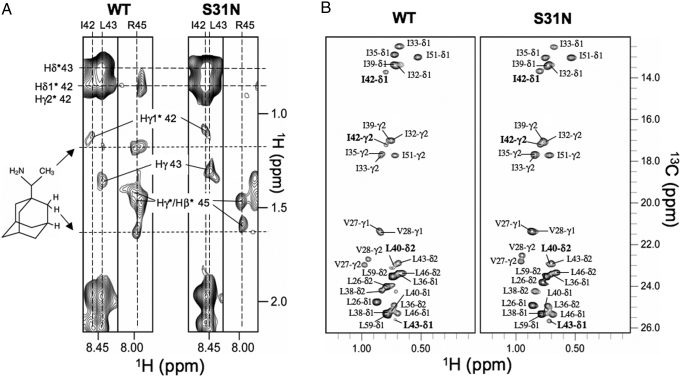
Fig. 4.
NMR investigation of rimantadine binding to the WT and S31N mutant. (A) Strips from 3D 15N-edited NOESY spectra (110 NOE mixing time) recorded for WT(18–60) and S31N(18–60) in the presence of rimantadine, indicating the absence of drug binding to the S31N mutant at the lipid-facing pocket. (B) WT(18–60) and S31N(18–60) methyl spectra show that the S31N mutation greatly reduced exchange broadening of the methyl-baring residues in the lipid-facing pocket.

