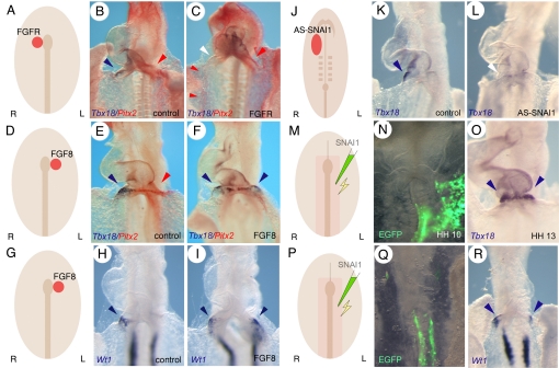
Fig. 3.
FGF8 and Snai1 are essential for right-sided PE development. (A, D, G, J, M, and P) Schematic depiction of the experiments. (B, E, H, and K) are controls and (C, F, I, L, N, O, Q, and R) experimental embryos. Only the heart region is shown in a ventral view, anterior end up of embryos subjected to whole mount in situ hybridization analysis of (B, C, E, and F) Pitx2 (red color) and Tbx18 (blue color), (H, I, R) Wt1, or (K, L, O) Tbx18 expression. (N and Q) GFP signal of electroporated embryos. (A-C) Embryos implanted with an aggregate of FGFR-expressing cells to the right of Hensen's node. (D-I) Embryos implanted with beads soaked in FGF8 protein to the left of Hensen's node. (J-L) Embryos treated with (K) control or (L) antisense Snai1 oligonucleotides. (M-R) Embryos electroporated with Snai1 and eGFP encoding plasmids. Blue arrowheads point to the endogenous and ectopic Tbx18 or Wt1 expression domains. Red arrowheads point to the endogenous and ectopic Pitx2 expression domains. White arrowheads in (C and L) point to the loss of endogenous Tbx18 expression.

