
Figure 1.
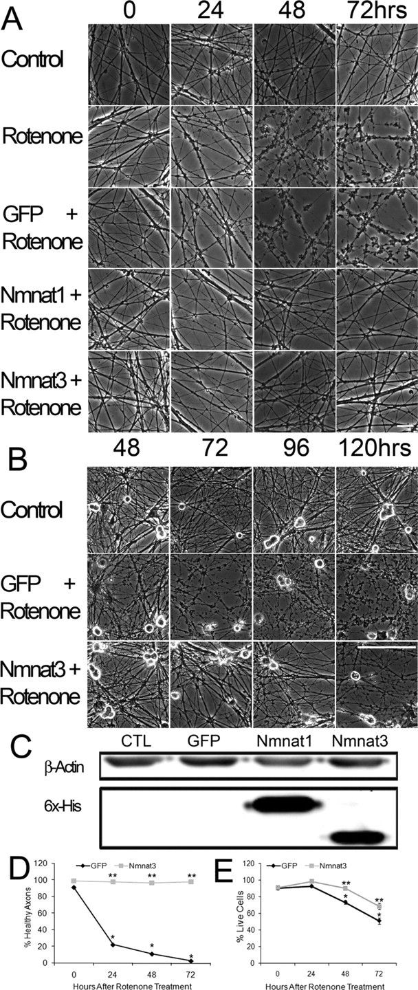
Nmnat expression protects against rotenone-mediated axonal degeneration in DRG neurons. A, B, DRG neurons were treated with rotenone (2.5 μm) for the indicated time, and phase-contrast microscopy was used to assess axonal degeneration. Degeneration is subtle after 24 h, but note the extensive axonal damage caused by rotenone by 48 h. Nmnat1- and Nmnat3-expressing neurons do not show axonal degenerative changes until after 96 h. Scale bars: A, 20 μm; B, 100 μm. C, Western-blot analysis of lysates of DRG neurons infected with lentivirus expressing GFP alone, Nmnat1, or Nmnat3 using a monoclonal antibody to the hexahistidine tag. D, Quantification of rotenone-induced axonal degeneration at 0, 24, 48, and 72 h in DRG neurons expressing GFP or Nmnat3 (4 fields per well, 6 wells per condition from duplicate experiments; see Materials and Methods). E, Quantification of surviving DRG neurons after treatment with rotenone for 24, 48, or 72 h using ethidium homodimer exclusion (6 wells per condition from duplicate experiments). *p < 0.01 compared with GFP-expressing neurons at 0 h; **p < 0.01 compared with rotenone-treated GFP-expressing neurons at corresponding time point. Error bars represent ±SEM.