
Figure 5.
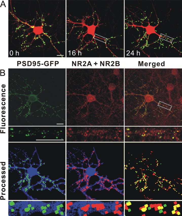
PSDs recovered after RAP-induced reversal of Tat-induced synapse loss contain NMDA receptor immunoreactivity. A, Representative images display labeled PSDs on a neuron expressing PSD95–GFP and DsRed2 before (0 h) and during (16 and 24 h) treatment with 50 ng/ml Tat. The LRP inhibitor RAP (50 nm) was added after 16 h. Eight hours after administering RAP, the number of synapses recovered (24 h frame). B, After collecting the live cell image at 24 h (as shown in A 24 h frame) the cells were fixed and labeled with antibodies to DsRed and the NR2A and 2B subunits of the NMDA receptor as described in Materials and Methods. Confocal micrographs display PSD95–GFP fluorescence (green) and NR2A and NR2B (red) immunoreactivity. Merged images display overlapping puncta (yellow). Processed images display puncta within the DsRed2 mask (blue). The same image-processing algorithm described in Materials and Methods was used to identify both NR2 immunoreactive puncta and PSD95–GFP puncta. The insets are enlarged images of the boxed region. Note that NR2A and NR2B immunoreactivity includes nontransfected cells in the field. Scale bar, 10 μm.