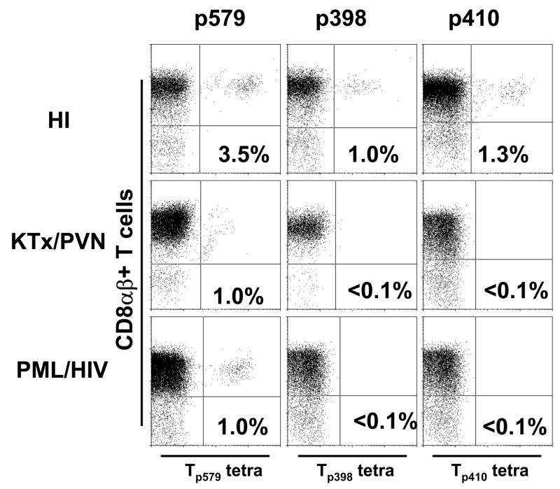
Figure 1.
Staining of PBMC from an HLA A*0201+ healthy individual, a PML patient and a KTx/PVN patient with tetrameric HLA-A*0201/BKV VP1p579, VP1p410 and VP1p398 complexes after in vitro stimulation with the respective peptides for 10–14 days. The percentages of CD8αβ+ T cells that bind the tetramers (dots in right upper quadrant of each panel) are indicated. Results were considered positive if the percentage of tetramer staining cells was equal or greater to 0.1% of CD8αβ+ T cells and formed a distinct population of cells on the dot plot.

