
Figure 6. Stably expressing MDA-MB-231 cell lines.
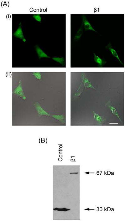
(A) Typical confocal XY (i) and merged bright-field images (ii) of MDA-MB-231 cells stably transfected with eGFP (‘Control’) and β1 with an eGFP C-terminal fusion (‘β1’). (B) Western blot of protein from MDA-MB-231 cells stably transfected with eGFP (‘Control’; total cell lysate) and β1 with an eGFP C-terminal fusion (‘β1’; membrane preparation) using an anti-GFP antibody. eGFP, 30 kDa; β1-eGFP, 67 kDa.