
Figure 8. Effects of β1 on morphology, adhesion, migration and proliferation of MDA-MB-231 cells.
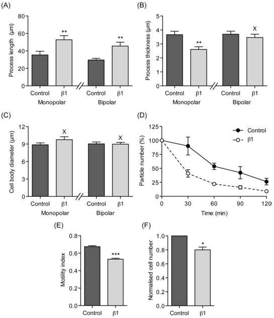
(A) Process length of MDA-MB-231 cells stably transfected with eGFP (‘Control’) and β1 with eGFP C-terminal fusion (‘β1’). (B) Process thickness of MDA-MB-231 cells stably transfected with eGFP (‘Control’) and β1 with eGFP C-terminal fusion (‘β1’). (C) Cell body diameter of MDA-MB-231 cells stably transfected with eGFP (‘Control’) and β1 with eGFP C-terminal fusion (‘β1’). In (A), (B) and (C), cells were defined as having monopolar or bipolar morphologies and analysed separately (n = 40). (D) Cell-cell adhesion. Single MDA-MB-231 cells stably transfected with eGFP (‘Control’) and β1 with eGFP C-terminal fusion (‘β1’) were incubated with gentle agitation for 2 h. The number of particles was monitored every 30 min and expressed as a percentage of the starting value. As cells adhered to one-another and formed aggregates, the particle number decreased (n = 3 repeat experiments). (E) Motility index (MI) of MDA-MB-231 cells stably transfected with eGFP (‘Control’) and β1 with eGFP C-terminal fusion (‘β1’) in a wound-heal assay. Wound width was measured at 0 h (W0) and 24 h (Wt). MI was calculated as 1 – (Wt/W0) (n = 135). (F) Proliferation of MDA-MB-231 cells stably transfected with eGFP (‘Control’) and β1 with eGFP C-terminal fusion (‘β1’), normalised relative to control. Cells were grown for 24 h and counted using the MTT assay (n = 3 repeat experiments). Data are presented as mean ± SEM. Significance: (X) P > 0.05; (*) P < 0.05, (**) P < 0.01, (***) P < 0.001; (A), (B), (C), (E) Student’s t-test; (F) Student’s paired t-test.