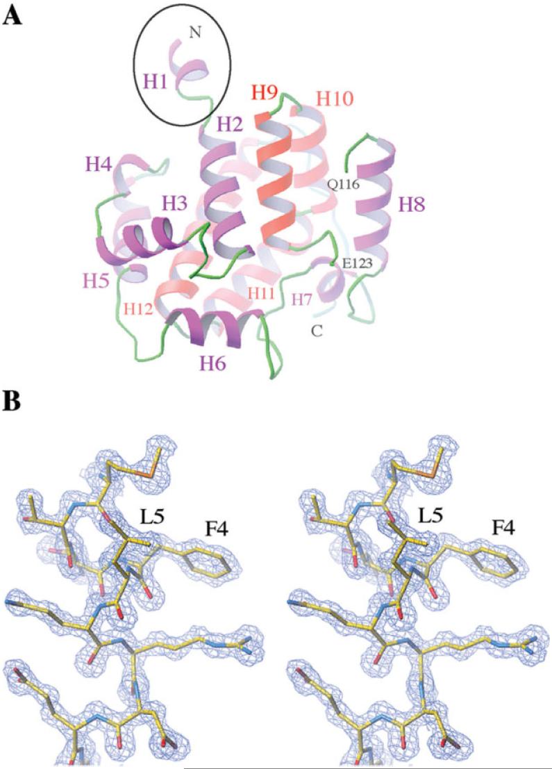
Figure 1.
(A) The all-alpha fold structure of DmsD from the S. typhimurium L12. The N-terminal extension is indicated by a black circle. The chain is broken at residues Q116 and E123 due to the missing residues. Helices from 1 to 12 are labeled. (B) Stereo view of the electron densities around the N-terminal extension (residues 1-8) at 1σ level. Residues are shown by stick representations.

