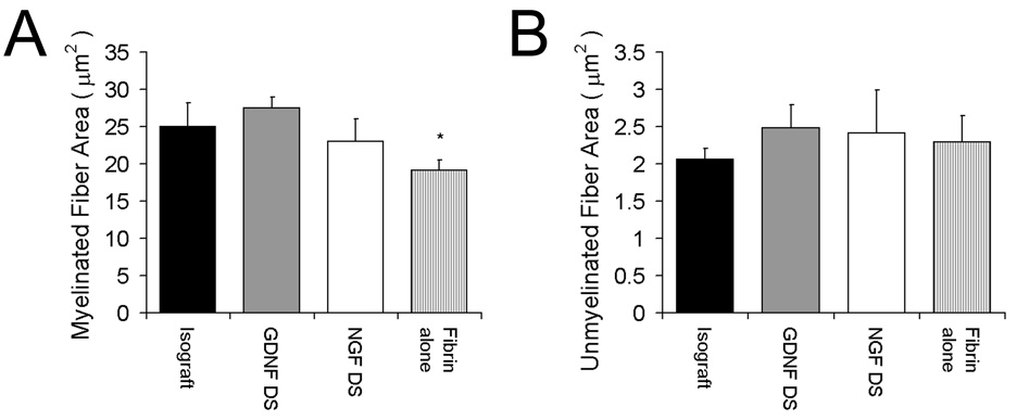
Fig. 6.
Myelinated and unmyelinated fiber areas of regenerating nerves at the midline of the conduit (or graft). The myelinated and unmyelinated fiber areas were determined from randomly selected specimens by electron microscopy from each group. The myelinated areas of for groups with the delivery system incorporating GDNF (GDNF DS) or NGF (NGF DS) were equivalent to the isograft, whereas the fibrin alone group had a lower myelinated fiber area (A). There were no differences among groups in unmyelinated fiber area (B). Data (n = 3) are shown as mean ± SEM. *Statistical significance (p < 0.05) compared to the isograft.

