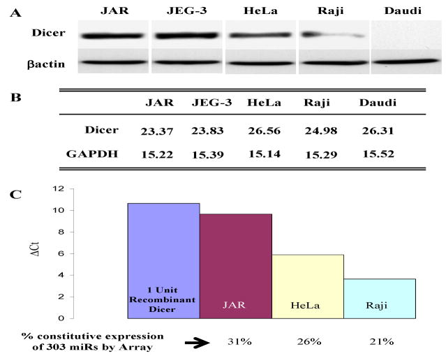
Figure 1. Dicer protein and activity are differentially regulated between cell types.
A. Untreated JAR, JEG-3, HeLa, Raji, and Daudi cells were analyzed by western blotting for Dicer and βactin levels. B. Dicer and GAPDH mRNA levels were assessed by quantitative real time RT-PCR for each of the above cell lines and are expressed as CT values. C. Dicer activity was assessed for JAR, HeLa, and Raji cells. Cytoplasmic extract from each cell line or recombinant Dicer enzyme was assayed with a synthetic pre-miR-122a and buffer at 37°C for 2 hours. Activity was determined by quantitative RT-PCR of mature miR-122a levels relative to the recombinant dicer and is presented as Δ CT values. Constitutively expressed miRs were determined by miRNA microarray analyses and are presented as the percentage of miRs constitutively expressed in each cell line.

