Figure 1.
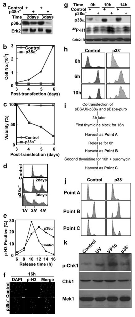
p38 MAP kinase is required for cell proliferation and survival. (a) HeLa cells were co-transfected with pBS/U6-p38 and pBabe-puro at a ratio of 9:1. After 1 day of incubation, puromycin was added for additional 1 or 2 days to select transfection-positive cells. After floating cells were removed, the remaining attached cells were harvested, and lysates were subjected to western blot using antibodies indicated on the left. (b, c) Cells were transfected and selected with puromycin as in (a). Cells were harvested at times as indicated, and cell proliferation (b) and viability (c) were monitored. To determine cell viability, floating cells and attached cells were harvested and counted separately. Viability was calculated as a percentage of attached cells compared to total cells. (d) Fluorescence-activated cell sorting (FACS) profiles of p38-depleted cells harvested at 2 days or 3 days post-transfection. (e) p38 was depleted in a synchronized culture using a double thymidine block protocol (16 h of thymidine block, 8 h of release and followed by a second 16 h incubation with thymidine). After release from the block for different times, cells were analysed by a phospho-histone H3 (p-H3) antibody staining. (f) Representative images of cells at 16 h post-release. Scale bar, 20 µm. (g) p38α was depleted in synchronized cells as in (e). After release from thymidine block for different times as indicated, cells were harvested and lysates were subjected to an anti-p38α immunoblot (top panel) or an anti-Cdc2 (middle panel) immunoprecipitation (IP)/kinase assay, using histone H1 as a substrate. (h) p38α was depleted in synchronized cells as in (e). After release from thymidine block for different times as indicated, cells were harvested for FACS analysis. (i) The detailed protocol to deplete p38α in well synchronized cells. (j) HeLa cells were depleted of p38a as in (i), harvested at three different time points as indicated and analysed by FACS. Point A, after first thymidine block for 16 h. Point B, after release for 8 h from the first thymidine block. Point C, after second thymidine block for 16 h. (k) HeLa cells were depleted of p38a as in (i), harvested at point C, and analysed by anti-phospho-Chk1 western blot to examine DNA damage checkpoint activation. As two positive controls, HeLa cells were either UV irradiated with 100 J/m2 and harvested after 1.5 h incubation (lane 2) or treated with 25 µg/ml VP16 for 4 h (lane 3).

