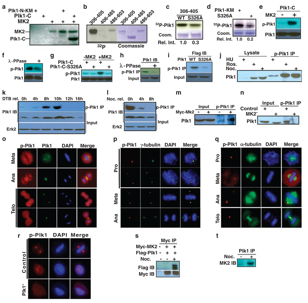
Figure 5.
MK2 targets Ser326 of Plk1. (a) Purified MK2 was incubated with Plk1 fragments in the presence of [γ-32P]ATP. The reaction mixtures were analysed by polyacrylamide gel electrophoresis, then autoradiography. Plk1-N-KM (kinase defective mutant): aa1–305; Plk1-C: aa306–603. (b) MK2 was incubated with three Plk1 C-terminal fragments (aa306–405, aa406–485 and aa486–603). (c, d) MK2 was incubated with Plk1-KM or a Ser326A mutant of different lengths (c, aa306–405; d, aa1–603). (e) MK2 was incubated with Plk1-C in the presence of cold ATP. The reaction mixtures were analysed by anti-phospho-Plk1 (p-Plk1) western blot. (f) After Plk1-C was incubated with MK2, the 50 µl reaction mixture was treated with 400 U λ-phosphatase (400 U µl−1; New England Biolab, Ipswich, MA, USA, Catalog number P0753) for 3 h at room temperature and analysed as in (e). (g) Purified Plk1-C (WT or S326A mutant) was incubated with MK2 and analysed by western blot. (h) HeLa cells were treated with nocodazole and harvested. After incubation with λ-phosphatase, lysates were subjected to anti-phospho-Plk1 immunoprecipitation (IP), followed by anti-Plk1 western blot. (i) HEK293 cells were transfected with Flag-Plk1 (WT or S326A mutant), and treated with nocodazole. Lysates were subjected to anti-phospho-Plk1 IP, followed by anti-Flag western blot. (j) HeLa cells were treated with hydroxyurea (lanes 1 and 4) or nocodazole (lanes 3 and 6) to block at S or M phase, respectively. To block cells at G2 phase, cells were released from the double thymidine block for 6 h, then incubated for 2 h in the presence of roscovitine (lanes 2 and 5). (k, l) HeLa cells were synchronized with the double thymidine block (k) or nocodazole treatment (l), released for the different times, and harvested. (m, n) HeLa cells were either MK2-overexpressed (m) or -depleted (n). (o–q) Mitotic cells were co-stained with a phospho-Plk1 antibody and antibodies against Plk1 (o), γ-tubulin (p) or α-tubulin (q). DNA was stained with DAPI. (r) Phospho-Plk1 staining is specific. Cells were Plk1-depleted with dsRNA and stained with phospho-Plk1 antibody. Scale bar, 5 µm. (s) HEK293 cells were co-transfected with Myc-MK2 and Flag-Plk1. At 1 day post-transfection, cells were treated with nocodazole for 12 h, and then harvested. Lysates were subjected to anti-Myc IP, followed by anti-Flag western blot analysis. (t) HeLa cells were treated with nocodazole, and harvested. Lysates were subjected to Plk1 IP, followed by anti-MK2 western blot analysis.

