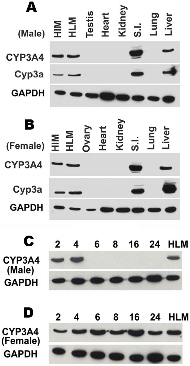
Figure 2. Tissue distribution and developmental expression patterns of CYP3A4 in TgCYP3A4/hPXR mice.
(A, B) Liver, lung, small intestine (S.I.), kidney, heat, testis/ovary were collected from transgenic male (A) and female (B) mouse at 4-week-old; (C, D) Liver tissues were collected from transgenic male (C) and female (D) mice of different ages (2-24 weeks). All microsomes were prepared by differential centrifugation. Pooled microsomal samples (3-4 in each group) were used for western blot analysis. The monoclonal antibody against CYP3A4 (mAb 275-1-2) recognizes human CYP3A4 but not mouse Cyp3a or other liver proteins. The monoclonal antibody against Cyp3a (mAb 2-13-1) reacts with both mouse Cyp3a and human CYP3A4. Human intestine microsomes (HIM) and human liver microsomes (HLM) served as positive controls for CYP3A4. GAPDH was used as loading control.

