
FIGURE 2.
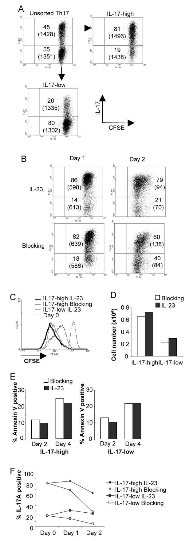
IL-23 maintains the IL-17 secreting phenotype without affecting cell expansion or survival. A, Naïve CD4+ T cells were activated, cultured in TGF-β+IL-6+IL-1β and blocking antibodies (anti-IFN-γ and anti-IL-4) for five days before sorting into IL-17-high and –low populations. Cells were then labeled with CFSE and for intracellular IL-17 following stimulation with PMA + ionomycin. Numbers indicate percent of cells in each quadrant and bracketed numbers indicate CFSE MFI. B, IL-17-high cells were cultured with IL-23 and blocking antibodies or blocking antibodies alone for the indicated times before cells were stimulated and stained for intracellular IL-17. Numbers indicate percent of cells in each quadrant and bracketed numbers indicate CFSE MFI. C, IL-17-high or -low CFSE-stained cells prepared as in B were cultured for two days with blocking antibodies in the presence or absence of IL-23 as indicated. CFSE staining is shown from freshly stained cells (Day 0) for comparison. D, IL-17-high and -low cells cultured as in B were counted after 48 hours. E, IL-17-high and -low cells were cultured as in B and were analyzed for Annexin V staining after two or four days of culture in the presence or absence of IL-23. F, IL-17-high and-low cells were stimulated with anti-CD3 and cultured with blocking antibodies with or without IL-23. Cells were stimulated with PMA+Ionomyocin for 4 hours and stained for intracellular IL-17.