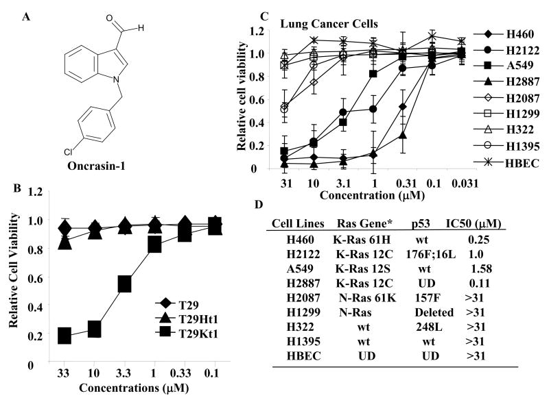
Figure 1.
Library screening. (A) Chemical structures of oncrasin-1. (B) Dose effect of oncrasin-1 in T29, T29Ht1, and T29Kt1 cells. The cells were treated with various concentrations (ranging from 0.1 μM to 33 μM) of oncrasin-1. Cell viability was determined 3 days after treatment by SRB assays. Control cells were treated with solvent (DMSO), and their value was set as 1. (C) Dose-Response in human lung cancer cell lines. Lung cancer cells with various oncogenic Ras gene status were treated with oncrasin-1 at various concentrations. Cell viability was determined as in B. The values shown are the means + SD of two assays done in quadruplicate. Control cells were treated with solvent (DMSO), whose value was set as 1. (D) The status of Ras gene and p53 gene mutations, and IC50 of oncrasin-1 in lung cancer cell lines tested in C. *Based on published data and the Cancer Genome Project Database (http://www.sanger.ac.uk/genetics/CGP/). Wt, wild type; UN, unknown; HBEC, human bronchial epithelial cells.

