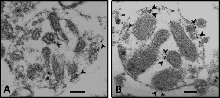
Figure 2.
Immunogold staining of εPKC in synaptosomes at 48 h of reperfusion after IPC. Electron micrographs showing synaptosomes isolated from hippocampus of sham (n = 3) and IPC-treated (n = 3) rats. A and B depict immunogold staining of εPKC in synaptosomes obtained from hippocampus of sham and IPC rat, respectively, at 48 h of reperfusion. The arrows depict immunogold/εPKC reactivity. Scale bar, 100 nm.

