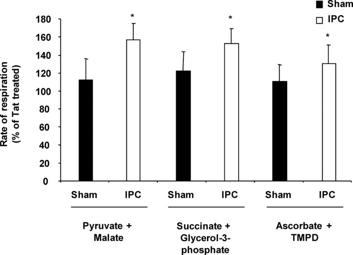
Figure 4.
Activation of εPKC increases the rate of mitochondrial respiration in synaptosomes isolated from hippocampus of ischemic preconditioned rats. Hippocampal synaptosomes were isolated at 48 h of reperfusion from sham (n = 6) or IPC-treated (n = 6) animals. This figure depicts substrate oxidation rates in hippocampal synaptosomes treated with tat or ψεRACK in both experimental groups. The results are expressed as mean ± SEM of percentage of oxygen consumption in tat-treated group. *p < 0.05, ψεRACK versus tat-treated synaptosomes.

