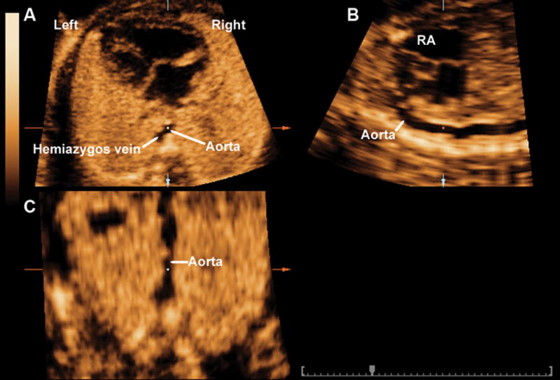
Figure 1.

Multiplanar display of the sagittal view of the ductal arch in a fetus with an interrupted left-sided inferior vena cava with hemiazygos continuation. The four-chamber view is displayed in panel A, in which a “two-vessel sign” can be seen, representing the hemiazygos vein to left of the aorta. The sagittal view of the ductal arch and the coronal view of the aorta are displayed in panels B and C, respectively. RA: right atrium.
