
Figure 2.
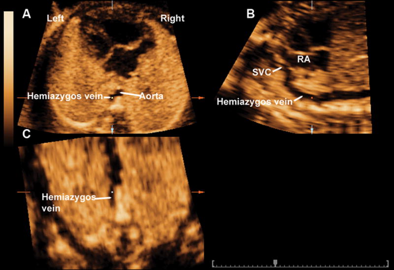
Multiplanar display of a dilated hemiazygos vein in a fetus with interrupted left-sided inferior vena cava with hemiazygos continuation. The reference dot was placed in the dilated hemiazygos vein in the four chamber view of the heart (panel A). This allowed for both the visualization of both the sagittal view of the hemiazygos vein in panel B and the coronal view of the dilated hemiazygos vein in panel C. Rotation of the coronal view to a vertical position on panel C allowed for visualization of the dilated hemiazygos vein draining into a persistent left SVC. SVC: superior vena cava; RA: right atrium.