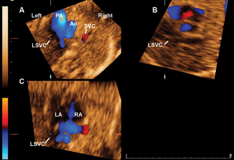
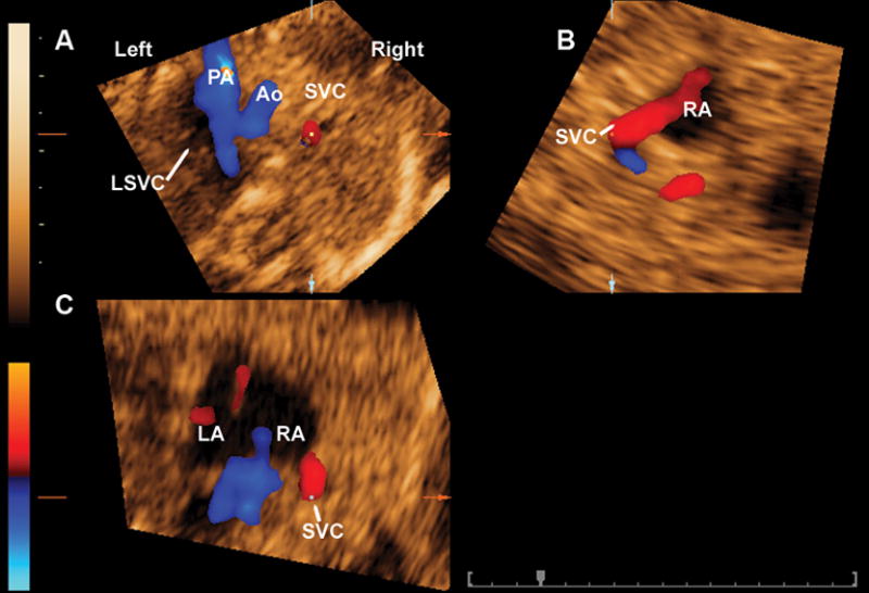
Figure 5.


A) Figure 5a shows the mutiplanar display of the three-vessel view of a fetus with persistent left SVC with dilated coronary sinus. The reference dot was placed on the vascular structure to the left of the pulmonary artery (panel A), which allowed for the clear visualization of a vascular structure (panel B) joining a dilated coronary sinus, which is projected into the left atrium (panel C). B) The same procedure was used to document the normal vascular connections of the SVC (Figure 5b). PA: pulmonary artery; Ao: aorta; LSVC: left superior vena cava; SVC: superior vena cava; RA; right atrium; LA: left atrium.
