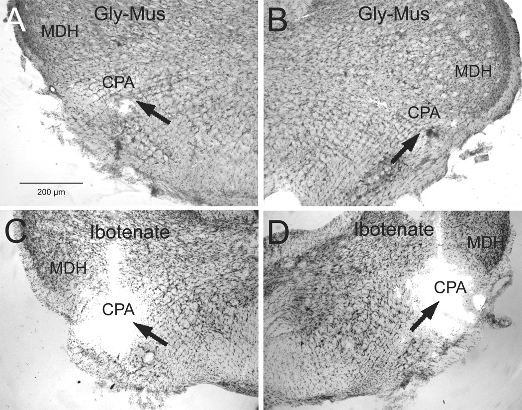
Figure 5.
Photomicrographs of typical injection sites (arrows) for deposits of glycine and muscimol (A, B; case 2218) and ibotenic acid (C, D; case 1559) into the most caudal ventrolateral medulla and centered in the CPA (arrows).Abbreviations: CPA, caudal pressor area; MDH, medullary dorsal horn.Line bar in A for figures A–D.

