Figure 2.
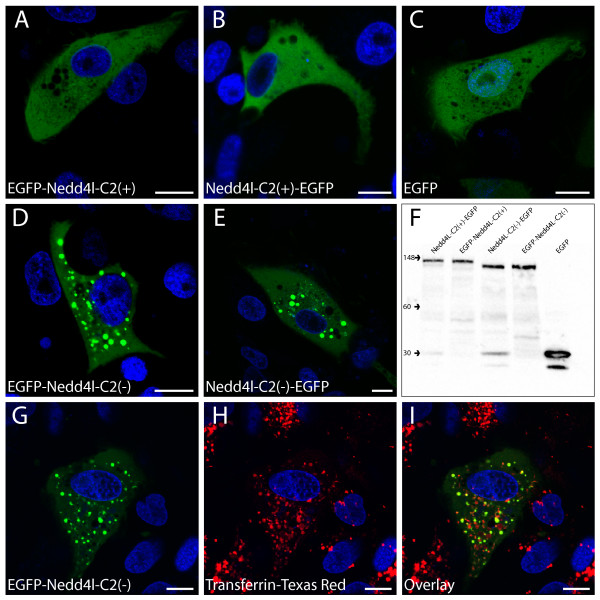
Differential subcellular localization of EGFP tagged NEDD4L-C2(+) and NEDD4L-C2(-) isoforms. Confocal microscopic images of X. laevis A6 cells transiently transfected with EGFP-NEDD4L-C2(+) (A), NEDD4L-C2(+)-EGFP (B), EGFP (C), EGFP-NEDD4L-C2(-) (D), and NEDD4L-C2(-)-EGFP (E). Western blot experiments of crude lysates from transiently transfected A6 cells (F) demonstrate that differential degradation of either isoform did not occur. The monoclonal EGFP antibody, JL-8 (Clontech) was used for detection. Confocal microscopic images of A6 cells transiently transfected with EGFP-NEDD4L-C2(-) and incubated with the early endosomal marker, Transferrin-Texas Red® (G-I) indicate that EGFP-NEDD4L-C2(-) localizes to the early endosome. Green (EGFP-tagged NEDD4L-C2(-)) and blue (nuclear marker) channels (G). Red (early endosome marker) and blue channels (H). Green, blue and red channel overlay (I). Scale bars are equivalent to 10 μm.

