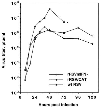
Figure 2.
Growth kinetics for rRSV/mIFN-γ, rRSV/CAT, and wt RSV in HEp-2 cells. Cell monolayers were infected with 2 pfu per cell (two replicate wells per virus), and 200 μl aliquots of supernatant were taken at the indicated time, adjusted to contain 100 mM magnesium sulfate and 50 mM Hepes buffer (pH 7.5), flash-frozen, and stored at −70°C until titration. Each aliquot taken was replaced with an equal amount of fresh medium. Each single-step growth curve represents the average of the virus titers from the two infected cell monolayers. The cell monolayer infected with wt RSV was more than 90% destroyed at 72 h postinfection (∗), whereas that infected with rRSV/mIFN-γ or rRSV/CAT was almost intact, reflecting attenuation of the chimeric virus in vitro.

