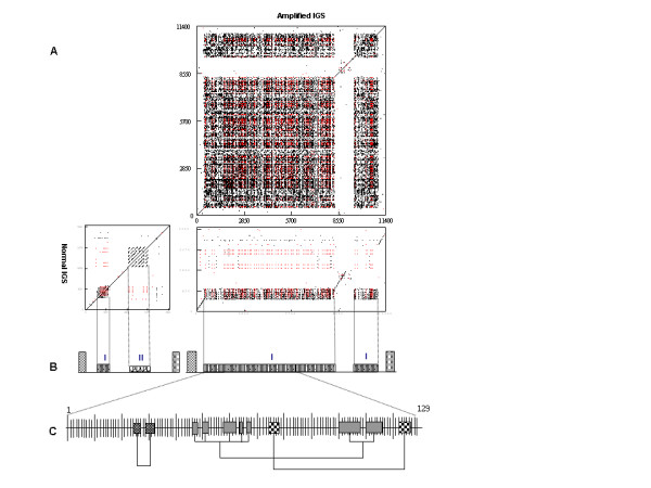
Figure 6.

Comparison of typical IGS and modified IGS. (A) Dot blot analysis of normal IGS from Sle0089P21 and amplified IGS from unit IV of HBa0007F24. Red dot represents complimentary match between two sequences. Dot blot parameter: window = 15, mismatch = 0. (B) Diagram representing the structures of normal IGS and amplified IGS of 25-18S rDNA. Type II subrepeat in the modified IGS is replaced with type I subrepeat. Left-side box represents 25S rDNA and right-side box represents 18S rDNA. (C) Fragmental duplication revealed by the Neighbor-joining tree among 129 monomers of IGS unit IV. The closely related pairs of monomers are connected.
