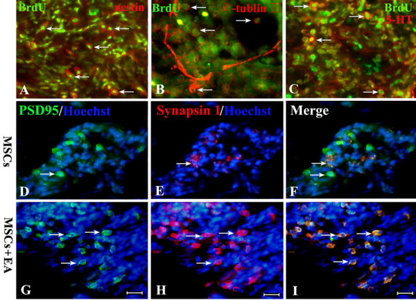
Figure 8.

Differentiation of MSCs into neuron-like cells at 8 weeks following transplantation. A, B and C: Using different makers (nestin for neuroblast cells (A), β-tublin III for young neurons (B), and 5-HT for functional neurons (C) combined with a BrdU double-label), we found that MSCs transplanted into injured spinal cord tissue could survive and differentiate into neuronal phenotypes in the MSCs+EA group. D-F: In the MSCs group, a double immunofluorescence study of PSD95 (green) and synapsin I (red) showed a small number of coexpressing cells. G-I: Double immunofluorescence showed that some cells coexpressed PSD95 (green) and synapsin I (red) in the grafts of the EA+MSCs group. Scale bars: 20 μm.
