Figure 2.

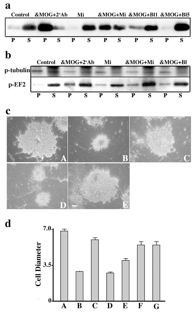
Fc Receptors mediate MOG cross-linking effects. Immunoblots show (a) MOG repartitioning into a TX-100 insoluble fraction (P) and (b) dephosphorylation of β-tubulin and increased phosphorylation of EF-2 in the pellet fraction, and (c) O4 staining shows process retraction (quantification of cell diameters shown in d) in OLs after treatment with anti-MOG 8-18C5 followed by either anti-mouse IgG (α-MOG+2°Ab, cB, dB) or microglia (α-MOG+Mi, cD, dD), but not after 30 min incubation with microglia alone (Mi, cC, dC) or anti-MOG plus microglia previously blocked with 1 (α-MOG+Bl1, dE), 5 (α-MOG+Bl5, cE, dF) or 10 (dG) μg/ml of anti-CD32/CD16. Under control conditions, MOG is mostly TX-100 soluble (S) and cells show normal morphology (cA, dA). Bar, 5 μm. dD vs. dE P < 0.05, dD vs. dF. p < 0.005 (Student’s t-test). The results shown are characteristic of three independent experiments.
