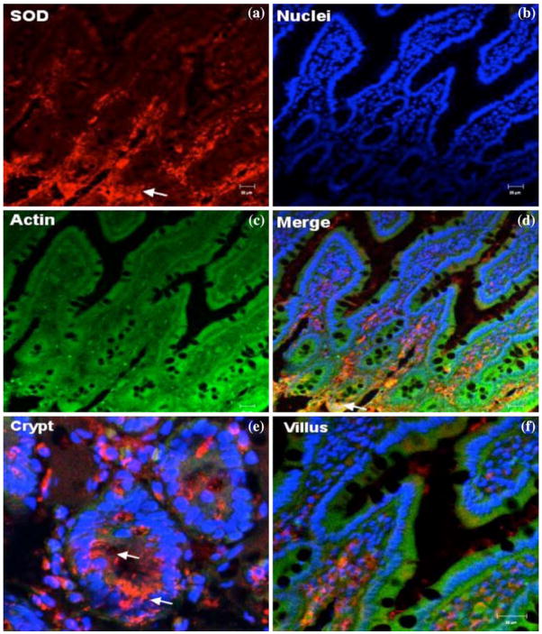
Fig. 3.
Confocal microscopic localization of SOD (red, a); Alexa Fluor 488 phalloidin (actin, green, c); Hoecht dye (Nuclei, blue, b) and an overlay merge image (d). Intenset immunoreactivity for SOD was observed in the crypt region, predominantly in the cytoplasm, while villus tip cells showed faint staining. Representative images of crypt base (e) and villus tip (f) regions are shown. Scale bars, 20 μm

