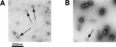
Figure 1.
Electron micrographs of L1 particles. After purification, particles were adsorbed to carbon-coated grids, were stained with 1% uranyl acetate, and were examined with a Philips electron microscope model EM 400RT at magnification ×36,000. (A) An L1-CCR5 particle preparation. Arrows identify putative 12-capsomere particles, which are ≈28 nm in diameter. (B) A preparation of wild-type L1 VLPs. Large 72-capsomere (55-nm) particles and a 12-capsomere (28-nm) particle (indicated by an arrow) are visible.

