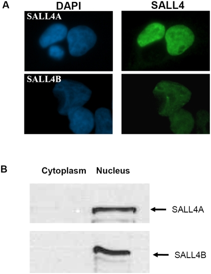
Figure 1. Nuclear localization of SALL4.
(A) Immunofluorescence of 293T cells transfected with either SALL4A (upper panel) or SALL4B (lower panel). Cells were stained with polyclonal anti SALL4 antibody (right) and DAPI (left). Both SALL4A and SALL4B were observed only in the nuclei of transfected. (B) Cellular fractionation with Western blotting showing SALL4A and SALL4B in the nuclei. Lysates from SALL4A or SALL4B-expressed 293Tcells were fractionated. 50 ug of subcellular fractions were separated on the SDS-PAGE and probed with the anti-SALL4 antibody.

