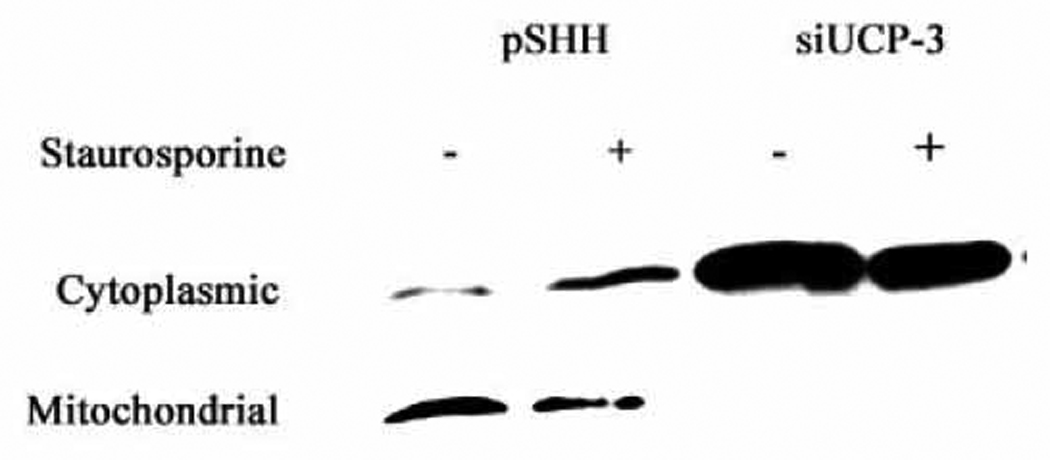
Figure 6. Cytochrome c release following an apoptogen challenge.

siUCP3 and pSHH (control) cells were challenged with 100 nM staurosporine for 6 h. Cytoplasmic and mitochondrial fractions were collected and subjected to Western blot analysis for cytochrome c localization. Note, the apoptogen caused release of cytochrome c from mitochondria to the cytosol in control cells. Also note the predominant localization of cytochrome c in the cytosolic fraction of the silenced chondrocytes, even in the absence of staurosporine.
