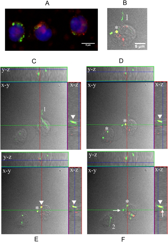
Figure 6. Isolated monocytes contain phagocytosed and degraded B. burgdorferi but not intact spirochetes.
(A) Epifluorescence image (100×) acquired from human monocytes incubated for 4-hours with Bb-GFP (MOI 100∶1) and labeled with the cell membrane marker FM4-64 (red) and the nuclear dye DAPI (blue). Phagocytosed (both coiled and degraded) Bb-GFP and fragmented nuclei were observed. (B–F) Orthogonal view (y,z and x,z axes) of optical sections (x,y axes) through a confocal stack of isolated monocytes incubated with Bb-GFP (MOI 100∶1) and labeled with lysotracker (red). (B) Digital enlargement of an extracellular spirochete (labeled # 1) in close proximity to a monocyte. Panels C–F are presented in 2 µm increments [(C) 6 µm, (D) 8 µm (E) 10 µm and (F) 12 µm] through the depth of the monocytes (total depth = 14 µm). Arrows and asterisks point to several internalized and degraded Bb-GFP, whereas colocalized phagolysosomes and fluorescent spirochetes shown in yellow are indicated by the arrowhead. The numbers 1 and 2 are placed next to individual extracellular Bb-GFP. Shapes within x,y axes are represented with their corresponding position in the x,z and y,z axes.

