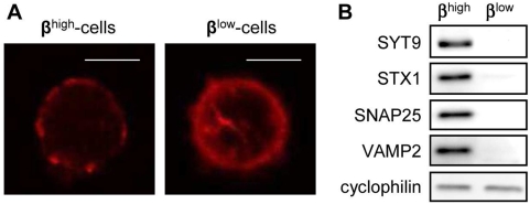
Figure 6. The exocytosis machinery of βlow-cells is not favorable to GSIS.
(A) Representative pictures of actin filament distribution in βhigh and βlow-cells. Scale bar: 10 µm (B) Representative immunoblots of exocytotic proteins in βhigh and βlow-cells (n = 3). n represents the number of independent cell preparations from at least 6 pooled rats each.

