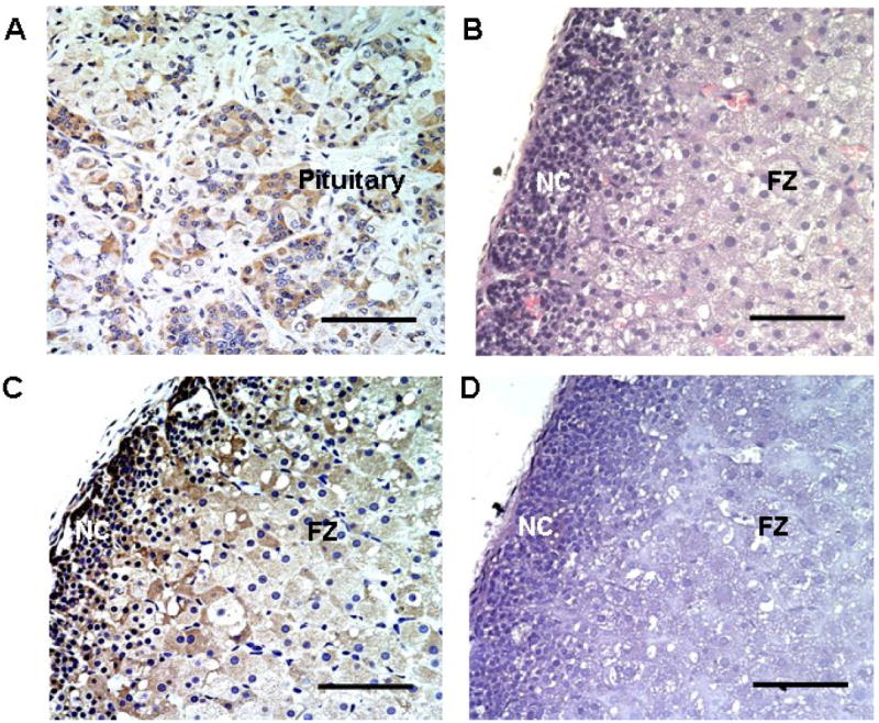
Fig. 4. Immunohistochemistry characterization of GnRHR expression in fetal adrenal.

Panel A. Positive control for GnRHR detection using human pituitary. Panel B. H&E staining of HFA showing both neocortex (NC) and fetal zone (FZ). Panel C. GnRHR staining in fetal adrenal gland. GnRHR protein (brown signal) was detectable in most of the neocortex and fetal zone cells. Panel D. Fetal adrenal processed for immunohistochemistry without inclusion of the GnRHR antibody (Negative control). All results are representative of staining from at least three fetal adrenals. Scale bar represents 10 μM in real size.
