
Fig. 5. ACTH and GnRH steroidogenic effects on fetal zone monolayer cultures.
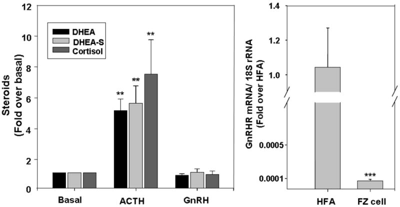
Panel A. Fetal zone cells were plated for 5 day before experiments. Cells were treated with ACTH (10 nM) or GnRH (10 nM) in 1 % serum for 24 h, and medium was collected for steroid assays. Data points are the mean ± SD of values from at least three independent experiments that are expressed as the fold change over basal (**, P < 0.01, compared to basal level). Panel B. Reduction of GnRHR mRNA expression in fetal zone cells after culturing. qPCR on GnRHR transcript was performed on cDNA from fetal adrenal tissues (HFA) or fetal zone cells in culture (FZ). The results were normalized to 18S rRNA as internal control. Data are expressed as fold over human fetal adrenal tissue level and are the mean ± SD of values from at least three different independent experiments (***, P<0.001).