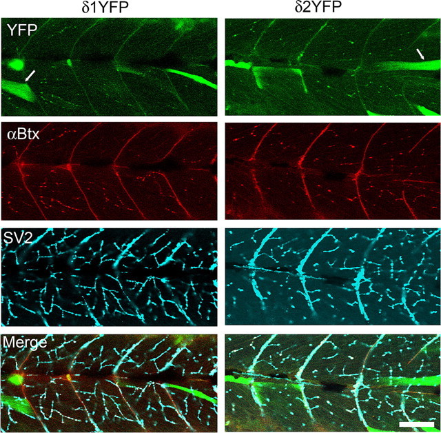
Figure 3.

Clustering of AChRs comprised of 2α, β, γ/ε and δ1YFP or δ2YFP subunits. Confocal images of sop−/− embryos expressing δ1YFP (left) or δ2YFP (right) were taken at 3dpf. Top panels show the YFP signal. Whereas some muscle cells with high expression of δ1/2YFP have cytoplasm filled with YFP signals (arrow), cells expressing lower level of δYFP exhibit clusters. These clusters of YFP colocalize with α-BTX signals (second row). In the third row, nerve terminals are visualized by anti-SV2 antibody. In muscle cells that express δ1/2YFP, YFP clusters, α-Btx and SV2 staining overlap and display white color in the merged picture (bottom). Scale bar, 50 μm.
