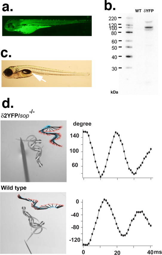
Figure 4.

a, YFP fluorescence of a 3dpf sop−/− embryo stably expressing δ2YFP (δ2YFP/sop−/−) in all muscle cells. b, Western blot of δ2YFP protein using antibody against YFP. A band is observed ∼110 kDa in δ2YFP/sop−/− embryos. A corresponding band is not present in wild-type fish. c, Lateral view of a δ2YFP/sop−/− larva at 7dpf. Notice the formation of a swim bladder (arrow). Sop−/− embryos without the transgene never form swim bladders. d, Movement of embryos at 3dpf in response to a tail stimulus. A δ2YFP/sop−/− embryo is shown in the top and a wild-type embryo (sop+/+ without the transgene) is shown in the bottom. Superimposed images of embryos at 0, 10, 20, 30, and 40 ms are shown to the left. Rostral midlines at each time point are shown as white lines. Insets represent swimming as a series of rostral midlines. The beginning of the line corresponding to the head is shown as red circles and the end of the line is shown as blue triangles. Head angles plotted against time for each trace are shown on the right. Note the initial angle is determined by the positioning of the embryo before touch and is different from trial to trial.
