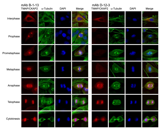
Figure 5.
Immunofluorescence staining showing the status of T596 phosphorylation at different stages of mitosis. C2C12 (mouse myoblast) cells were fixed and immunostained for TMAP/CKAP2 (Cy3; red) and α-tubulin (AlexaFluor 488; green) using mAb B-1-13 (left panel) and mAb D-12-3 (right panel). DAPI (blue) staining shows nuclei and chromosomes. mAb B-1-13 produced the staining patterns which were consistent with previous reports (left panels). Although mAb D-12-3 also successfully detected microtubule-associated TMAP/CKAP2 in interphase cells, it failed to detect TMAP/CKAP2 in mitotic cells at prophase, prometaphase, and metaphase (right panels). However, the expected staining patterns returned again starting from the anaphase and were indistinguishable to those produced by mAb B-1-13.

