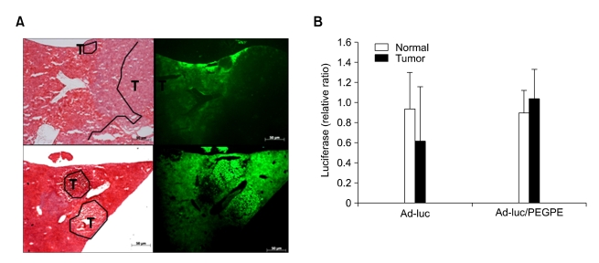
Figure 4.
Expression of liposome-complexed adenovirus in liver metastasis tumor model. (A) GFP expression in intrahepatic tumors (T) and adjacent liver following systemic administration of adenoviruses. 1 × 10¹¹ vp of Ad-GFP or Ad-GFP/PEGPE was injected intravenously and livers, including tumors were removed after 24 h. Eight-µm frozen sections were prepared and mounted with cover glass. Fluorescence was observed by Olympus fluorescent microscope (×40). After then, the slides were stained with hematoxylin and eosin, and observed under the light microscope (×40). (B) Relative ratio of luciferase expression of intrahepatic tumors to adjacent non-neoplastic liver following systemic administration of adenoviruses. 1 × 10¹¹ vp of Ad-luc or Ad-luc/PEGPE was intravenously injected and luciferase expression was measured in tumors and adjacent normal livers. Luciferase activity in tumors was calculated by relative to that in adjacent non-neoplastic liver. Data represents the mean ± S.D.

