Figure 1.
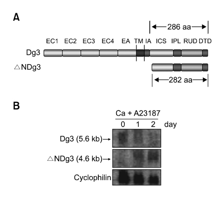
(A) Overall structure of Dg3 and ΔNDg3. EC1-EC4, four extracellular cadherin-typical repeats; EA, extracellular anchor domain; TM, transmembrane domain; IA, intracellular anchor domain; ICS, intracellular cadherin-specific domain; IPL, proline-rich linker domain; RUD, repeating unit domain; DTD, desmoglein-specific terminal domain. ΔNDg3 contains slightly shorter ICS, IPL, RUD and DTD domains. (B) Northern blot analysis. HaCaT cells were treated with 1 µM A23187 and 0.3 mM calcium for the indicated time points. About 5.6 kb Dg3 mRNA and 4.6 kb ΔNDg3 mRNA were shown. Cyclophilin was detected as a loading control.

